
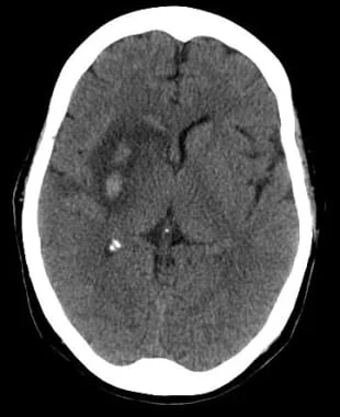

Abstract
Haemorrhagic stroke is a neurological emergency caused by the rupture of a cerebral blood vessel, resulting in bleeding within the brain parenchyma or the subarachnoid space. It is associated with high mortality, significant neurological morbidity and substantial long-term disability. The two principal forms are intracerebral haemorrhage (ICH) and subarachnoid haemorrhage (SAH), each characterised by distinct aetiologies, clinical manifestations and management strategies. Rapid recognition, urgent neuroimaging, physiological stabilisation and targeted treatment are essential to prevent secondary brain injury and improve outcomes. This review discusses the background, aetiology, pathophysiology, epidemiology, clinical presentation, diagnostic approach, acute treatment, complications and prognosis of haemorrhagic stroke.
Keywords: Haemorrhagic stroke; intracerebral haemorrhage; subarachnoid haemorrhage; cerebral aneurysm; hypertension; neurocritical care; stroke management.
1. Overview
1.1 Background and definition
Stroke is a leading cause of death and acquired neurological disability worldwide. It is conventionally classified into ischaemic stroke, caused by interruption of cerebral blood flow, and haemorrhagic stroke, caused by bleeding from a ruptured blood vessel.
Haemorrhagic stroke represents approximately 10–20% of all strokes in many high-income populations, although its proportion varies considerably by geography and population characteristics. Despite being less common than ischaemic stroke, it contributes disproportionately to early mortality and severe disability.
The two major forms are:
Intracerebral haemorrhage (ICH): Bleeding directly into the brain parenchyma, producing a haematoma that damages surrounding tissue. It may occur in deep structures, lobar regions, the cerebellum or the brainstem.
Subarachnoid haemorrhage (SAH): Bleeding into the subarachnoid space between the arachnoid and pia mater. Aneurysmal rupture is a major cause of spontaneous SAH and can result in abrupt increases in intracranial pressure and subsequent cerebral ischaemia.
Other forms of intracranial bleeding include subdural and epidural haemorrhage. These are usually associated with trauma and are not generally classified as spontaneous haemorrhagic stroke.
Distinguishing the type of haemorrhage is essential because the underlying pathology, diagnostic investigations and treatment priorities differ.
1.2 Aetiology
The causes of haemorrhagic stroke are heterogeneous and depend on the patient’s age, vascular risk factors, medication exposure and underlying cerebral pathology.
Hypertensive arteriopathy
Chronic hypertension is a major risk factor for spontaneous intracerebral haemorrhage. Prolonged exposure to elevated arterial pressure damages small penetrating arteries, particularly those supplying the basal ganglia, thalamus, pons and cerebellum.
Small-vessel degeneration and rupture may result in bleeding into these deep brain structures. Hypertensive haemorrhage can also occur in other locations.
Cerebral amyloid angiopathy
Cerebral amyloid angiopathy (CAA) results from the deposition of amyloid-beta protein within cortical and leptomeningeal blood vessel walls. This weakens the vessels and increases their susceptibility to rupture.
CAA is an important cause of lobar intracerebral haemorrhage in older adults and may be associated with recurrent haemorrhage, cortical superficial siderosis and multiple cerebral microbleeds.
Intracranial aneurysms
An intracranial aneurysm is a localised dilation of an artery caused by structural weakness of its wall. Rupture allows arterial blood to enter the subarachnoid space.
Aneurysmal SAH is associated with considerable early mortality and a high risk of rebleeding if the aneurysm is not secured promptly.
Vascular malformations
Arteriovenous malformations, cavernous malformations and other vascular abnormalities can predispose patients to intracranial bleeding. These causes are particularly relevant in younger individuals without established hypertensive or degenerative vascular disease.
Coagulation disorders and antithrombotic medication
Anticoagulants, thrombocytopenia, coagulation factor deficiencies and other haemostatic abnormalities can increase the risk and severity of intracranial haemorrhage.
Anticoagulant-associated ICH is particularly concerning because bleeding may continue or expand unless the anticoagulant effect is reversed appropriately.
Other causes
Less common causes include intracranial neoplasms, haemorrhagic transformation of cerebral infarction, cerebral venous thrombosis, vasculopathies and exposure to sympathomimetic drugs such as cocaine or amphetamines.
Traumatic brain injury is another important cause of intracranial haemorrhage, although traumatic bleeding is distinguished from spontaneous haemorrhagic stroke in most clinical classifications.
1.3 Risk factors
Risk factors can be divided into modifiable and non-modifiable categories.
Modifiable risk factors
- Uncontrolled hypertension.
- Cigarette smoking.
- Excessive alcohol consumption.
- Cocaine and amphetamine use.
- Anticoagulant exposure and inappropriate antithrombotic use.
- Poorly controlled vascular risk factors.
- In selected populations, untreated vascular abnormalities.
Non-modifiable or less readily modifiable risk factors
- Increasing age.
- Genetic predisposition to aneurysm formation or vascular disease.
- Family history of intracranial aneurysm or aneurysmal SAH.
- Cerebral amyloid angiopathy.
- Certain inherited vascular disorders.
Hypertension is among the most important preventable contributors to intracerebral haemorrhage. Risk reduction therefore depends heavily on sustained blood pressure control and appropriate management of other vascular risk factors.
1.4 Pathophysiology
The pathophysiology of haemorrhagic stroke involves both the initial mechanical injury caused by bleeding and subsequent secondary processes that may enlarge the area of brain damage.
Primary brain injury
Following vessel rupture, blood enters the brain tissue or subarachnoid space. In ICH, the accumulating haematoma compresses adjacent structures and disrupts neuronal and glial tissue.
The extent of primary injury depends on the volume and location of the haemorrhage. Bleeding in strategically important structures, such as the brainstem, may cause severe deficits even when the haematoma is relatively small.
In SAH, blood spreads through the cerebrospinal fluid spaces and may increase intracranial pressure abruptly. This can reduce cerebral perfusion and, in severe cases, produce transient global cerebral ischaemia.
Haematoma expansion
In ICH, bleeding may continue after the initial vessel rupture. Haematoma expansion is associated with neurological deterioration and poorer functional outcomes.
The risk is influenced by factors such as early presentation, elevated blood pressure, anticoagulant use and imaging markers of ongoing bleeding. This explains the importance of prompt assessment, blood pressure management and anticoagulation reversal when indicated.
Perihaematomal oedema and inflammation
Blood products trigger inflammatory responses and biochemical cascades that contribute to secondary tissue injury.
Thrombin activation, haemoglobin breakdown, iron accumulation, oxidative stress and disruption of the blood–brain barrier contribute to perihaematomal oedema. This may increase local tissue pressure and exacerbate neuronal dysfunction.
Increased intracranial pressure
Intracranial haemorrhage increases the volume of intracranial contents. Because the skull is a rigid compartment, compensatory mechanisms eventually become insufficient.
Rising intracranial pressure reduces cerebral perfusion pressure. Severe pressure elevation can cause tissue displacement, brainstem compression and cerebral herniation.
Intraventricular extension of an intracerebral haematoma may obstruct cerebrospinal fluid circulation, causing acute hydrocephalus.
Secondary injury in subarachnoid haemorrhage
After aneurysmal SAH, several processes contribute to delayed neurological deterioration:
- Cerebral vasospasm, involving narrowing of cerebral arteries.
- Delayed cerebral ischaemia.
- Microcirculatory dysfunction and impaired autoregulation.
- Inflammatory and thrombotic mechanisms.
- Hydrocephalus and disturbances in cerebrospinal fluid circulation.
Vasospasm and delayed cerebral ischaemia are related but distinct phenomena; delayed ischaemia may occur through mechanisms beyond large-vessel narrowing alone.
These processes explain why patients with SAH require prolonged neurological monitoring even after the aneurysm has been secured.
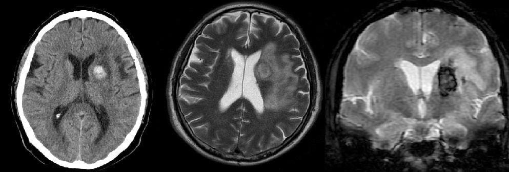
1.5 Epidemiology
Haemorrhagic stroke accounts for a smaller proportion of all stroke events than ischaemic stroke, but its clinical burden is substantial.
Incidence and outcomes vary according to geographical region, age distribution, hypertension prevalence, access to emergency care and availability of specialist stroke services.
Intracerebral haemorrhage is strongly associated with hypertension, small-vessel disease and cerebral amyloid angiopathy. Deep haemorrhages are commonly linked to hypertensive arteriopathy, whereas lobar haemorrhages in older adults may suggest CAA.
Aneurysmal SAH occurs less frequently than ICH but often affects adults during working age. Smoking, hypertension and family history are recognised risk factors.
Improvements in emergency imaging, neurocritical care, aneurysm treatment and rehabilitation have enhanced aspects of care, but haemorrhagic stroke remains associated with substantial mortality and disability.
1.6 Prognosis
Prognosis is determined by the severity and location of the haemorrhage, the initial neurological state, the presence of intraventricular bleeding, hydrocephalus, haematoma expansion, age, comorbidities and treatment delays.
For ICH, large haematomas, brainstem involvement, reduced consciousness and intraventricular extension are associated with worse outcomes.
For SAH, prognosis is influenced by the initial neurological grade, the volume and distribution of blood, rebleeding, hydrocephalus and delayed cerebral ischaemia.
Survivors may experience persistent motor impairment, language difficulties, cognitive dysfunction, fatigue, depression, anxiety and reduced independence. Recovery may continue over months or years, depending on the nature and extent of the initial injury.
Prognostic scores can support clinical assessment but should not be used in isolation to determine treatment limitation or individual outcomes.
2. Clinical Presentation
The clinical presentation of haemorrhagic stroke depends on the anatomical location, volume and rate of bleeding. Symptoms may develop abruptly, progress over minutes to hours, or fluctuate as complications emerge.
2.1 General clinical features
Common manifestations include:
- Sudden focal weakness or paralysis.
- Facial asymmetry.
- Speech disturbance, including dysarthria or aphasia.
- Sensory loss.
- Headache.
- Nausea and vomiting.
- Visual disturbance.
- Ataxia, vertigo or impaired coordination.
- Confusion or altered consciousness.
- Seizures.
Headache, vomiting and reduced consciousness may be prominent in haemorrhagic stroke, but their absence does not exclude intracerebral bleeding.
The clinical presentation alone cannot reliably distinguish haemorrhagic stroke from ischaemic stroke. Urgent brain imaging is therefore essential.
2.2 Clinical presentation of intracerebral haemorrhage
ICH commonly produces focal neurological deficits that reflect the affected anatomical region.
Basal ganglia haemorrhage: May cause contralateral weakness, sensory deficits and gaze abnormalities.
Thalamic haemorrhage: May produce prominent sensory impairment, altered consciousness and sometimes ocular movement abnormalities.
Lobar haemorrhage: May present with aphasia, neglect, visual field deficits, seizures or focal weakness, depending on the cortical region involved.
Cerebellar haemorrhage: May cause headache, vomiting, vertigo, gait instability, dysarthria and impaired coordination. Deterioration may result from brainstem compression or obstructive hydrocephalus.
Brainstem haemorrhage: May cause profound impairment of consciousness, cranial nerve abnormalities, limb weakness and respiratory or cardiovascular instability.
A rapid decline in consciousness should raise concern about haematoma expansion, increasing intracranial pressure, intraventricular extension or hydrocephalus.
2.3 Clinical presentation of subarachnoid haemorrhage
Aneurysmal SAH classically presents with a thunderclap headache: a sudden, severe headache reaching maximum intensity rapidly.
Additional features include:
- Neck pain or stiffness.
- Photophobia.
- Nausea and vomiting.
- Brief or prolonged loss of consciousness.
- Seizures.
- Focal neurological deficits.
- Confusion or reduced consciousness.
Some patients experience a preceding headache, sometimes described as a sentinel headache. Such episodes are not specific, but a sudden severe headache should never be dismissed without appropriate assessment.
SAH may initially present without clear focal neurological deficits. A normal initial neurological examination does not reliably exclude the diagnosis.
2.4 Clinical assessment and severity
Initial assessment should include the Glasgow Coma Scale, pupillary examination, assessment of focal deficits and documentation of vital signs.
The NIH Stroke Scale can help quantify neurological deficits, although it may underestimate some important features of haemorrhagic stroke.
In ICH, the ICH Score is commonly used as a prognostic aid. In aneurysmal SAH, the Hunt and Hess and World Federation of Neurosurgical Societies grading systems help describe clinical severity.
These scales support communication and risk assessment but must be interpreted alongside imaging, clinical trajectory and the patient’s broader circumstances.
3. Diagnosis
The objectives of diagnostic assessment are to confirm the haemorrhage, establish its location and extent, identify the underlying cause, detect complications and guide urgent treatment.
3.1 Initial emergency assessment
Management begins with assessment and stabilisation of airway, breathing and circulation.
Important early steps include:
- Establishing symptom onset or last-known-well time.
- Assessing consciousness and neurological deficits.
- Measuring blood pressure, oxygen saturation, temperature and blood glucose.
- Establishing intravenous access.
- Reviewing anticoagulants, antiplatelet agents and relevant medical history.
- Arranging urgent brain imaging.
Airway protection and ventilatory support may be required in patients with severe impairment of consciousness.
3.2 Non-contrast computed tomography
Non-contrast CT of the head is usually the first-line imaging investigation for suspected acute intracranial haemorrhage.
It can rapidly identify acute blood and establish the anatomical distribution of the haemorrhage. It also helps detect mass effect, intraventricular extension, hydrocephalus and midline shift.
In ICH, CT findings help estimate haematoma volume and identify features associated with expansion. Serial imaging may be indicated when clinical deterioration occurs or when expansion is suspected.
3.3 CT angiography
CT angiography is used when an underlying vascular lesion is suspected or when additional information may influence management.
In SAH, CT angiography is important for detecting an intracranial aneurysm and planning treatment.
In ICH, vascular imaging may help identify aneurysms, arteriovenous malformations, other vascular lesions or imaging markers associated with ongoing bleeding.
The decision to undertake angiography depends on the haemorrhage pattern, age, clinical history and initial imaging findings.
3.4 Magnetic resonance imaging
MRI may provide additional information when the underlying cause remains uncertain.
Susceptibility-weighted imaging and gradient-echo sequences can detect cerebral microbleeds and other evidence of previous bleeding. MRI can help identify cerebral amyloid angiopathy, cavernous malformations, haemorrhagic tumours and selected cases of haemorrhagic transformation.
MRI is generally not the initial investigation when immediate CT is available and acute haemorrhage must be excluded rapidly.
3.5 Diagnosis of subarachnoid haemorrhage
If a patient presents with a suspected SAH, urgent non-contrast CT is required.
Diagnostic accuracy is highest when CT is performed early after symptom onset. If CT is negative, further investigation depends on the timing of imaging, the quality and interpretation of the scan, the clinical presentation and local guidance.
Under NICE guidance, routine lumbar puncture is not recommended when a CT performed within six hours of symptom onset, and appropriately reported, is negative. If CT is performed more than six hours after onset and is negative, lumbar puncture should be considered when clinically indicated. When undertaken to investigate SAH, it should generally occur at least 12 hours after symptom onset to assess for bilirubin (xanthochromia) using the recommended laboratory method.
A confirmed SAH should prompt urgent vascular imaging, usually CT angiography, to identify the bleeding source. Digital subtraction angiography may be required if the cause remains uncertain. <Cite refs={[“turn702359search2”]}/>
3.6 Laboratory investigations
Laboratory testing supports the assessment of bleeding risk, physiological disturbances and treatment eligibility.
Typical investigations include:
- Full blood count and platelet count.
- Prothrombin time and international normalised ratio.
- Activated partial thromboplastin time when indicated.
- Renal function and electrolytes.
- Blood glucose.
- Liver function when clinically relevant.
- Blood group and compatibility testing when transfusion may be required.
Specific coagulation tests may be needed in patients receiving direct oral anticoagulants, depending on the agent and local laboratory capability.
3.7 Differential diagnosis
Important differential diagnoses include:
- Acute ischaemic stroke.
- Cerebral venous thrombosis.
- Seizure with postictal neurological deficits.
- Hypoglycaemia.
- Migraine with neurological manifestations.
- Reversible cerebral vasoconstriction syndrome.
- Other causes of thunderclap headache.
- Intracranial infection or mass lesions.
Rapid imaging is essential because therapies appropriate for ischaemic stroke, particularly intravenous thrombolysis, may cause serious harm when intracranial haemorrhage is present.
4. Treatment and Management
Treatment should be initiated immediately and adapted to the haemorrhage type, severity, cause and associated complications. Patients with significant haemorrhage should receive specialist stroke, neurocritical care or neurosurgical assessment, with transfer to an appropriate centre when necessary.
4.1 Initial stabilisation
Initial priorities include:
- Maintaining airway patency and adequate ventilation.
- Supporting circulation and avoiding hypotension.
- Correcting hypoglycaemia and clinically significant metabolic disturbances.
- Providing oxygen when hypoxaemia is present.
- Monitoring neurological status and vital signs.
- Preventing aspiration and other complications of immobility.
Intubation may be necessary in patients who cannot protect their airway or who develop respiratory failure.
Analgesia and antiemetics may be required. Sedating medications should be used carefully because they can interfere with neurological assessment.
4.2 Blood pressure management in intracerebral haemorrhage
Elevated blood pressure is common after ICH and may be associated with haematoma expansion.
In selected patients with mild-to-moderate spontaneous ICH, controlled early reduction of systolic blood pressure may improve functional outcomes and reduce haematoma expansion. Treatment should aim for smooth, sustained control rather than large fluctuations.
NICE recommends considering rapid blood pressure lowering in appropriate patients presenting within six hours with systolic pressure between 150 and 220 mmHg, with a target of 140 mmHg or lower and a reduction not exceeding 60 mmHg in the first hour. These recommendations do not apply universally; patients with major haemorrhage, very low consciousness scores, structural vascular causes or planned early surgery require individualised specialist decisions. <Cite refs={[“turn702359search1”]}/>
Intravenous antihypertensive agents may be selected according to local protocols and patient-specific factors.
The aim is to balance the potential benefit of limiting further bleeding against the risk of reducing cerebral perfusion.
4.3 Reversal of anticoagulation
Anticoagulant-associated ICH requires urgent assessment of the medication, time of last dose, renal function and coagulation status.
- Warfarin: Intravenous vitamin K and four-factor prothrombin complex concentrate are generally used for rapid reversal.
- Dabigatran: Idarucizumab is a specific reversal agent when indicated and available.
- Factor Xa inhibitors: Andexanet alfa may be appropriate in selected patients receiving apixaban or rivaroxaban, while prothrombin complex concentrate may be used under relevant protocols.
- Other coagulation abnormalities: Management is directed at the underlying defect and the clinical context.
The benefits and risks of reversal agents, including thrombosis, must be considered. Treatment should follow current local protocols and specialist advice. <Cite refs={[“turn702359search4″,”turn702359search5”]}/>
4.4 Neurosurgical treatment of intracerebral haemorrhage
Surgical intervention is considered according to the location and volume of the haematoma, neurological deterioration, mass effect and presence of hydrocephalus.
Potential interventions include haematoma evacuation, minimally invasive procedures in selected cases, external ventricular drainage for hydrocephalus and decompressive surgery in carefully selected patients.
Cerebellar haemorrhage requires particular attention because relatively small increases in volume can compress the brainstem or obstruct cerebrospinal fluid circulation.
Not all intracerebral haemorrhages benefit from evacuation. Decisions should be made by an experienced multidisciplinary team, considering the evidence, the patient’s condition and the goals of care.
4.5 Treatment of aneurysmal subarachnoid haemorrhage
The principal objectives are to prevent rebleeding, preserve cerebral perfusion and manage delayed complications.
Securing the aneurysm
The ruptured aneurysm should be treated as early as feasible, preferably within 24 hours when possible. Treatment generally involves one of two approaches:
- Endovascular coiling, in which coils are introduced into the aneurysm to promote thrombosis.
- Microsurgical clipping, in which a clip is placed across the aneurysm neck.
The preferred technique depends on aneurysm anatomy, patient factors, available expertise and the likelihood of achieving durable occlusion. <Cite refs={[“turn702359search0″,”turn702359search2”]}/>
Nimodipine
Enteral nimodipine is recommended in aneurysmal SAH because it reduces the risk of poor neurological outcomes associated with delayed cerebral ischaemia.
Blood pressure and other physiological variables must be monitored, particularly if hypotension occurs.
Management of hydrocephalus
Acute hydrocephalus may require external ventricular drainage to relieve pressure and restore cerebrospinal fluid circulation.
Management of delayed cerebral ischaemia
Patients require neurological monitoring during the period of risk. Symptomatic delayed cerebral ischaemia may require specialist-directed interventions to improve cerebral perfusion.
Routine prophylactic hypervolaemia or indiscriminate haemodynamic augmentation is not recommended because of the risk of iatrogenic complications. <Cite refs={[“turn702359search0″,”turn702359search6”]}/>
4.6 Intracranial pressure management
Raised intracranial pressure may result from a large haematoma, cerebral oedema, hydrocephalus or obstructed cerebrospinal fluid circulation.
Management may include head positioning, adequate analgesia and sedation, correction of physiological disturbances, hyperosmolar therapy when indicated, cerebrospinal fluid drainage and selected surgical interventions.
Mechanical ventilation and advanced intracranial pressure monitoring may be required in critically ill patients.
Treatment must be individualised because interventions that reduce intracranial pressure can also affect systemic blood pressure and cerebral perfusion.
4.7 Seizure management
Seizures may occur after ICH or SAH. Clinical seizures should be treated promptly.
Continuous electroencephalographic monitoring may be appropriate when unexplained impaired consciousness raises suspicion of non-convulsive seizures.
Routine prophylactic antiseizure medication is not indicated for every patient with spontaneous ICH or SAH. The decision should reflect the haemorrhage characteristics, clinical events and relevant specialist guidance.
4.8 General supportive care
Supportive care is essential for reducing complications and improving recovery.
Swallowing and nutrition: Patients should undergo swallowing assessment before oral food, fluids or medication are given when dysphagia is suspected. Alternative nutrition may be required when swallowing is unsafe.
Venous thromboembolism prevention: Intermittent pneumatic compression is recommended for appropriate immobile patients. The timing of pharmacological prophylaxis depends on haemorrhage stability, repeat imaging and individual bleeding and thrombosis risks.
Temperature and glucose: Fever and significant glucose disturbances should be identified and treated.
Infection prevention: Appropriate respiratory care, aspiration prevention, skin care and catheter management help reduce complications.
Early rehabilitation: Rehabilitation should begin when medically appropriate, with the timing and intensity tailored to the patient’s neurological status and stability.
4.9 Rehabilitation and long-term management
Recovery after haemorrhagic stroke requires a coordinated multidisciplinary approach.
Rehabilitation may involve physiotherapy, occupational therapy, speech and language therapy, neuropsychology, dietetics and specialist nursing.
Long-term follow-up should address functional recovery, cognitive impairment, mood disorders, fatigue, swallowing difficulties and social participation.
Secondary prevention includes blood pressure control, smoking cessation, moderation of alcohol consumption and treatment of relevant vascular risk factors.
Restarting anticoagulants or antiplatelet agents after haemorrhagic stroke requires an individualised assessment of the indication for treatment, recurrent bleeding risk and thromboembolic risk.
After aneurysmal SAH, follow-up vascular imaging may be necessary to assess aneurysm occlusion, detect recurrence and identify other aneurysms when appropriate.
5. Complications
Complications may occur during the acute phase or develop during recovery.
5.1 Early complications
Haematoma expansion: Continued bleeding may increase tissue damage and worsen neurological deficits.
Raised intracranial pressure: Increasing pressure can impair cerebral perfusion and cause herniation.
Hydrocephalus: Blood may obstruct cerebrospinal fluid pathways, particularly after intraventricular haemorrhage or SAH.
Seizures: Seizures may occur at presentation or during the hospital course.
Rebleeding: Particularly important in aneurysmal SAH before the aneurysm is secured.
Cardiopulmonary complications: Severe brain injury may be accompanied by arrhythmias, myocardial injury, respiratory failure and aspiration pneumonia.
5.2 Delayed complications
Delayed complications include cerebral vasospasm and delayed cerebral ischaemia after SAH, venous thromboembolism, infection, contractures, pressure injuries, depression, anxiety and cognitive impairment.
Patients may experience persistent problems with memory, attention, executive function, language and emotional regulation even when motor recovery is relatively good.
Early recognition and targeted rehabilitation can improve quality of life and functional independence.
6. Prognosis and Outcomes
The prognosis of haemorrhagic stroke varies considerably.
6.1 Factors associated with outcome
Important predictors include:
- Initial neurological status.
- Haematoma volume and location.
- Intraventricular extension.
- Hydrocephalus and mass effect.
- Haematoma expansion.
- Age and pre-existing comorbidities.
- Anticoagulant-associated bleeding.
- Rebleeding or delayed cerebral ischaemia after SAH.
- Time to diagnosis and access to specialist treatment.
Prognostic scores can support communication and clinical decision-making, but they cannot predict an individual patient’s recovery with certainty.
6.2 Mortality and functional recovery
ICH carries a high risk of early death and long-term dependency, particularly when the haemorrhage is large or involves critical structures.
Aneurysmal SAH also carries a substantial risk of death and disability, including among patients who survive the initial bleeding event.
Recovery depends on the severity of the initial injury, secondary complications and the intensity and appropriateness of rehabilitation.
Some patients regain substantial independence, whereas others require prolonged support or ongoing assistance with daily activities.
6.3 Long-term quality of life
Long-term effects may include physical disability, fatigue, cognitive impairment, depression, anxiety and changes in social functioning.
Follow-up should therefore extend beyond motor recovery to include cognition, psychological health, communication, participation and caregiver needs.
7. Prevention
Prevention strategies focus on reducing the risk of vessel rupture and managing underlying vascular disease.
Key interventions include:
- Effective long-term hypertension management.
- Smoking cessation.
- Avoidance of cocaine and other illicit sympathomimetic drugs.
- Moderation of alcohol consumption.
- Appropriate prescribing and monitoring of anticoagulants.
- Evaluation of suspected intracranial aneurysms and vascular malformations.
- Individualised follow-up for patients with previous intracranial haemorrhage.
Screening for unruptured intracranial aneurysms is not routinely indicated for the entire population. It may be considered in selected higher-risk groups following specialist assessment.
8. Conclusion
Haemorrhagic stroke is a life-threatening cerebrovascular emergency characterised by bleeding into the brain parenchyma or subarachnoid space. Hypertension, cerebral amyloid angiopathy, intracranial aneurysms, vascular malformations and coagulation abnormalities are important underlying causes.
Clinical presentation varies according to the haemorrhage type and anatomical location. Rapid neurological assessment and urgent CT imaging are essential for diagnosis, while angiographic imaging and MRI may help identify the underlying cause.
Management requires a targeted approach incorporating physiological stabilisation, appropriate blood pressure control, anticoagulation reversal, neurosurgical or endovascular treatment when indicated, and prevention of secondary complications.
Long-term outcomes depend on the initial injury, complications and rehabilitation. Effective prevention, coordinated specialist care and comprehensive follow-up are central to reducing mortality, disability and recurrent vascular events.
Clinical take-home messages
- Haemorrhagic stroke must be distinguished rapidly from ischaemic stroke because treatment strategies differ.
- Non-contrast CT is the usual first-line investigation for suspected acute intracranial haemorrhage.
- Blood pressure reduction and anticoagulation reversal should be guided by the haemorrhage type, severity and current protocols.
- Aneurysmal SAH requires urgent identification and treatment of the ruptured aneurysm.
- Hydrocephalus, raised intracranial pressure, seizures and delayed cerebral ischaemia require active surveillance.
- Rehabilitation and long-term vascular risk management are essential components of care.
Further reading and clinical references
The expanded article should be checked against current clinical guidance before publication. These are the most useful authoritative references for the diagnostic and treatment sections:
- Medscape — original article supplied.
- AHA/ASA — 2022 Guideline for the Management of Patients With Spontaneous Intracerebral Hemorrhage.
Stroke
- AHA/ASA — 2023 Guideline for the Management of Patients With Aneurysmal Subarachnoid Hemorrhage.
- NICE — Stroke and transient ischaemic attack in over 16s: diagnosis and initial management.
NICE
- NICE — Subarachnoid haemorrhage caused by a ruptured aneurysm: diagnosis and management.
