

Abstract
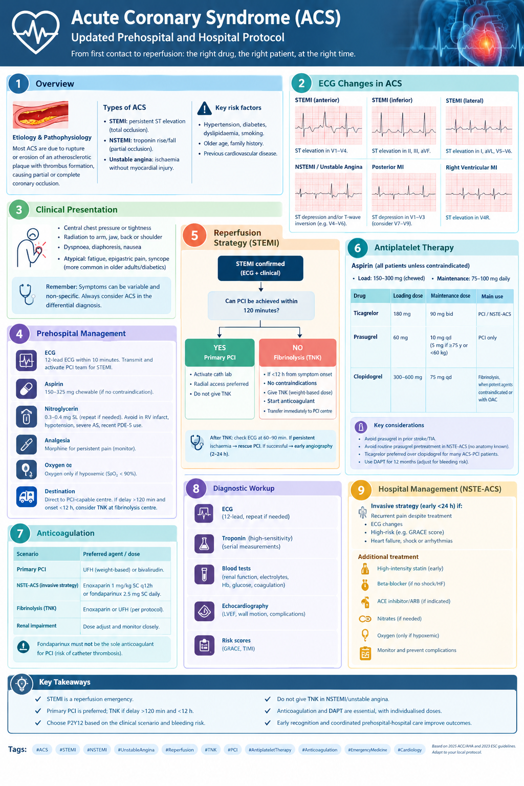
Acute coronary syndrome (ACS) is a spectrum of clinical conditions caused by acute myocardial ischaemia, most commonly resulting from disruption of an atherosclerotic coronary plaque and subsequent thrombus formation. It includes ST-segment elevation myocardial infarction (STEMI), non-ST-segment elevation myocardial infarction (NSTEMI), and unstable angina.
Early recognition, rapid electrocardiographic assessment, appropriate antithrombotic treatment and timely coronary reperfusion are essential to reduce mortality and preserve myocardial function. The 12-lead electrocardiogram (ECG) remains the principal initial investigation for identifying patients who require immediate reperfusion, while high-sensitivity cardiac troponin (hs-cTn) enables the diagnosis and risk stratification of patients without persistent ST-segment elevation.
This review discusses the pathophysiology, clinical presentation, ECG interpretation, diagnostic strategy and contemporary management of ACS, with particular emphasis on the decisions faced by emergency and prehospital clinicians. It includes practical recommendations for antiplatelet therapy, anticoagulation, primary percutaneous coronary intervention (PCI) and fibrinolysis with tenecteplase.
Keywords: acute coronary syndrome, STEMI, NSTEMI, ECG, myocardial infarction, primary PCI, tenecteplase, fibrinolysis, antiplatelet therapy, emergency medicine.
1. Introduction and pathophysiology
Acute coronary syndrome is a clinical manifestation of acute myocardial ischaemia caused by an imbalance between myocardial oxygen supply and demand. In the majority of cases, the underlying mechanism is atherosclerotic coronary artery disease complicated by plaque rupture or erosion and the formation of an intraluminal thrombus.
Plaque disruption exposes thrombogenic material to circulating blood, activating platelets and the coagulation cascade. The resulting thrombus may partially or completely obstruct coronary blood flow. Distal embolisation, vasoconstriction and microvascular dysfunction may further compromise myocardial perfusion.
The extent of myocardial injury depends on several interacting factors, including the location and severity of the obstruction, the duration of ischaemia, collateral circulation, myocardial oxygen demand and the effectiveness of reperfusion.
Persistent coronary occlusion can produce progressive myocardial necrosis. Early reperfusion limits infarct size, preserves left ventricular function and reduces the risk of heart failure, malignant ventricular arrhythmias and death.
Although plaque rupture is the most common mechanism, ACS can also occur through plaque erosion, coronary artery dissection, coronary embolism, epicardial coronary spasm or other mechanisms. Myocardial infarction with non-obstructive coronary arteries (MINOCA) is an important working diagnosis when infarction is established but angiography does not demonstrate obstructive coronary disease; it requires further investigation rather than being considered a final aetiological diagnosis.
1.1 Classification of acute coronary syndromes
ACS is classified according to the clinical presentation, ECG findings and cardiac biomarkers.
ST-segment elevation myocardial infarction (STEMI) is characterised by persistent ST-segment elevation or an accepted equivalent pattern consistent with acute coronary occlusion. It requires immediate assessment for reperfusion.
Non-ST-segment elevation myocardial infarction (NSTEMI) is diagnosed when there is acute myocardial injury, demonstrated by a rise or fall in cardiac troponin with at least one value above the assay-specific 99th percentile, together with clinical evidence of acute myocardial ischaemia. The ECG may show ST-segment depression, T-wave inversion, transient ST elevation or no diagnostic abnormalities.
Unstable angina describes acute myocardial ischaemia without evidence of acute myocardial injury sufficient to meet the definition of myocardial infarction. Its diagnosis has become less frequent with the widespread use of hs-cTn assays.
The distinction between STEMI and NSTE-ACS is operationally important because it influences the urgency and type of reperfusion strategy. It does not imply that NSTEMI is necessarily a less dangerous condition: patients with NSTE-ACS may develop cardiogenic shock, malignant arrhythmias or extensive myocardial injury.
1.2 Epidemiology and risk factors
Coronary artery disease remains a major cause of morbidity and mortality worldwide. The probability of ACS increases with age and the accumulation of cardiovascular risk factors.
Important modifiable risk factors include:
- Arterial hypertension.
- Diabetes mellitus.
- Dyslipidaemia.
- Cigarette smoking.
- Obesity and physical inactivity.
- Chronic kidney disease.
- Unhealthy dietary patterns.
Non-modifiable or historical risk factors include increasing age, family history of premature coronary disease and established atherosclerotic cardiovascular disease.
Clinicians should maintain a high index of suspicion in patients with diabetes, older adults, women and patients with chronic kidney disease, because presentations may be atypical or symptoms may be less prominent than expected.
1.3 Prognosis
Prognosis depends on the extent of myocardial damage, the time to reperfusion, left ventricular function, haemodynamic stability, renal function and associated comorbidities.
The early phase carries a substantial risk of ventricular tachycardia, ventricular fibrillation, acute heart failure, cardiogenic shock and mechanical complications. Later complications include adverse ventricular remodelling, chronic heart failure, recurrent ischaemia and recurrent cardiovascular events.
Rapid diagnosis and coordinated prehospital and hospital management are therefore central to improving outcomes.
2. Clinical presentation
2.1 Typical symptoms
The classic presentation is retrosternal pressure, heaviness, tightness or discomfort, often lasting more than 10–20 minutes. The pain may radiate to one or both arms, the shoulders, neck, jaw, back or epigastrium.
Associated symptoms include:
- Dyspnoea.
- Diaphoresis.
- Nausea or vomiting.
- Pallor and cold clammy skin.
- Weakness, light-headedness or syncope.
- A sense of impending death.
Symptoms may occur at rest or with minimal exertion. Relief after nitrates does not confirm ACS, and lack of response does not exclude it.
2.2 Atypical presentations
Some patients present without chest pain. Dyspnoea, acute pulmonary oedema, syncope, unexplained weakness, epigastric discomfort or acute confusion may be the predominant manifestations.
Atypical presentations are particularly relevant in older adults, patients with diabetes, women and people with chronic kidney disease. These presentations should not be dismissed as non-cardiac solely because the pain is absent or non-classical.
2.3 Initial clinical assessment
The first assessment should rapidly establish whether the patient is haemodynamically stable and whether there is evidence of ongoing myocardial ischaemia or a life-threatening complication.
Assess:
- Airway, breathing and circulation.
- Heart rate, blood pressure, respiratory rate, oxygen saturation and temperature.
- Level of consciousness and peripheral perfusion.
- Signs of acute heart failure, pulmonary oedema or cardiogenic shock.
- Pulse asymmetry, new aortic regurgitation murmur or other features raising suspicion of acute aortic syndrome.
- Relevant contraindications to antithrombotic treatment or fibrinolysis.
Important alternative diagnoses include acute aortic syndrome, pulmonary embolism, pericarditis, myocarditis, pneumothorax and oesophageal pathology. A differential diagnosis should be pursued when clinically indicated, but additional testing must not unnecessarily delay reperfusion in a patient with a convincing acute coronary occlusion pattern.
3. ECG interpretation in acute coronary syndrome
3.1 Timing and acquisition
A 12-lead ECG should be acquired and interpreted within 10 minutes of first medical contact or initial presentation. This is a time-critical recommendation in contemporary ACS guidance.
If the initial ECG is non-diagnostic but symptoms persist, recur or worsen, repeat ECGs should be obtained. A single normal ECG cannot exclude ACS. ECG changes may evolve over minutes, and comparison with previous tracings can be extremely valuable.
Continuous rhythm monitoring is appropriate in suspected high-risk ACS. Additional leads should be recorded when the clinical picture or standard ECG suggests posterior or right ventricular involvement.
3.2 Criteria for ST-segment elevation
In the appropriate clinical context, new ST-segment elevation measured at the J point in at least two anatomically contiguous leads supports the diagnosis of STEMI.
The usual thresholds are:
- At least 1 mm in leads other than V2–V3.
- In V2–V3: at least 2.5 mm in men younger than 40 years, 2 mm in men aged 40 years or older, and 1.5 mm in women.
These thresholds are aids to interpretation, not a substitute for clinical judgement. Important exceptions and mimics include left ventricular hypertrophy, bundle branch block, ventricular pacing, early repolarisation, pericarditis, myocarditis, Brugada pattern and Takotsubo syndrome.
A new left bundle branch block (LBBB) alone is not sufficient to diagnose acute coronary occlusion. In patients with LBBB or ventricular pacing, appropriate criteria such as the modified Sgarbossa criteria may help identify an acute occlusion pattern.
3.3 ECG patterns by infarction territory
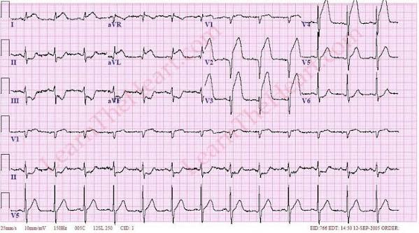
A. Anterior and anteroseptal STEMI
Typical leads: V1–V4, sometimes extending to V5–V6, I and aVL.
Likely culprit: left anterior descending coronary artery (LAD).
ST elevation in V1–V4 suggests anteroseptal or anterior injury. More extensive ST elevation extending into the lateral leads may indicate a larger territory at risk. Reciprocal ST depression may be present in the inferior leads.
A proximal LAD occlusion may produce extensive anterior changes, haemodynamic compromise and a high risk of left ventricular dysfunction. New conduction abnormalities and ventricular arrhythmias may occur.
Clinical interpretation should consider the distribution and magnitude of ST elevation, reciprocal changes, haemodynamic status and the evolution of the ECG.
Example ECG:
B. Inferior STEMI
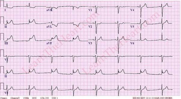
Typical leads: II, III and aVF.
Likely culprit: right coronary artery (RCA) more commonly, or left circumflex coronary artery (LCx), depending on coronary anatomy.
Inferior STEMI often produces reciprocal ST depression in leads I and aVL. ST elevation greater in lead III than in lead II may suggest RCA occlusion, particularly when accompanied by other right coronary territory findings, but it is not definitive on its own.
Inferior infarction may be complicated by sinus bradycardia, atrioventricular block, hypotension or right ventricular infarction. Assess for haemodynamic instability and obtain right-sided leads when right ventricular involvement is suspected.
Nitrates require particular caution when right ventricular infarction or hypotension is suspected because these patients may be preload-dependent.
Example ECG:
C. Lateral STEMI
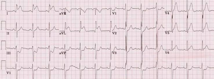
Typical leads: I, aVL, V5 and V6.
Likely culprit: LCx, diagonal branches of the LAD or other lateral wall branches.
Lateral infarction may be isolated or associated with anterior or inferior infarction. High-lateral infarction may be subtle, with changes confined predominantly to I and aVL.
Reciprocal ST depression in the inferior leads may strengthen suspicion when the lateral changes are small. Small areas of lateral infarction can be missed if the ECG is interpreted only by looking for marked ST elevation in the precordial leads.
Example ECG:
D. Posterior myocardial infarction
Posterior infarction is particularly important because it may not produce conventional ST elevation on the standard 12-lead ECG.
The standard ECG may show:
- Horizontal ST depression in V1–V3.
- Relatively tall R waves in V1–V3.
- Upright terminal T waves in the affected anterior leads.
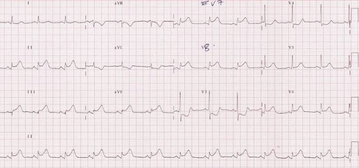
These findings may represent the reciprocal image of posterior ST elevation. When posterior infarction is suspected, record leads V7–V9. ST elevation in the posterior leads supports posterior STEMI; the usual threshold is lower than in standard limb and precordial leads, commonly at least 0.5 mm in two contiguous posterior leads, with higher thresholds sometimes applied in younger men.
Posterior infarction may accompany inferior or lateral STEMI or occur in association with LCx occlusion. Persistent symptoms with suggestive anterior ST depression should not be classified automatically as NSTEMI without considering posterior occlusion.
Example ECG and explanation:
E. Right ventricular myocardial infarction
Right ventricular infarction frequently accompanies inferior STEMI, especially with proximal RCA occlusion.
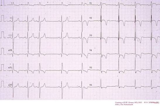
The standard ECG may show inferior ST elevation and ST elevation in V1. When right ventricular involvement is suspected, obtain right-sided leads, particularly V3R and V4R. ST elevation in V4R is a useful finding in the appropriate setting.
The clinical picture may include hypotension, elevated jugular venous pressure and relatively clear lung fields. This combination should prompt urgent assessment for right ventricular infarction, although not every patient exhibits the complete triad.
Management implications include careful fluid assessment and avoidance of unnecessary preload reduction. Nitrates and diuretics may worsen hypotension in a preload-dependent patient. Fluids should be administered cautiously, guided by clinical status and evidence of congestion, rather than given automatically.
Example ECG:
F. NSTEMI and subendocardial ischaemia
NSTEMI may produce horizontal or down-sloping ST depression, T-wave inversion, transient ST elevation or non-specific changes. Some patients have a normal initial ECG.
Dynamic ST depression in multiple leads may indicate widespread subendocardial ischaemia, particularly when accompanied by ongoing pain, haemodynamic instability or elevated troponin.
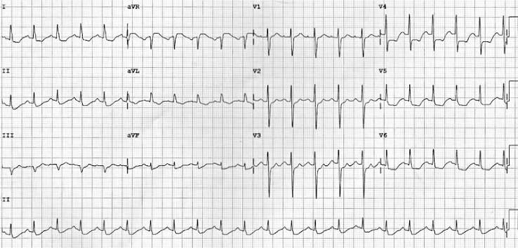
Diffuse ST depression with ST elevation in aVR can be a marker of severe global subendocardial ischaemia and may occur with left main, multivessel or proximal LAD disease. It is not specific for left main occlusion and must be interpreted in context, including possible supply–demand imbalance.
The absence of ST elevation does not indicate a benign condition. The urgency of invasive assessment should be determined by the clinical risk profile and evidence of ongoing ischaemia.
Example ECG:
G. De Winter pattern and other occlusion patterns
The De Winter pattern consists of upsloping ST depression at the J point in the precordial leads, followed by tall, symmetrical T waves. It is associated with acute coronary occlusion, often involving the proximal LAD, and should prompt urgent specialist assessment for reperfusion.
Other concerning patterns include hyperacute T waves, transient ST elevation, posterior infarction patterns, and selected cases with LBBB or ventricular pacing in which modified Sgarbossa criteria suggest acute coronary occlusion.
These patterns reinforce an important principle: coronary occlusion cannot be excluded simply because standard STEMI thresholds are not met.
3.4 ECG changes over time
The ECG may evolve from hyperacute T waves to ST elevation, followed by the appearance of pathological Q waves and T-wave inversion. The exact sequence and degree of these changes vary; Q waves do not always develop, and reperfusion may alter the expected evolution.
An ECG obtained after pain resolves may look different from the tracing recorded during active ischaemia. Serial recordings should therefore be interpreted in relation to symptom onset, treatment and clinical changes.
3.5 Practical ECG interpretation checklist
For each ECG, systematically assess:
- Rhythm and heart rate.
- ST-segment elevation or depression.
- T-wave morphology and symmetry.
- Anatomical distribution and contiguous leads.
- Reciprocal changes.
- QRS duration, bundle branch block and pacing.
- Evidence of posterior or right ventricular involvement.
- Comparison with previous and subsequent ECGs.
The ECG should be integrated with the history, examination, haemodynamic state and biomarkers. Neither an isolated ECG feature nor a single normal tracing should be used in isolation to exclude ACS.
4. Diagnostic investigations
4.1 High-sensitivity cardiac troponin
Hs-cTn is the preferred biomarker for detecting myocardial injury. Diagnosis of acute myocardial infarction requires a rise and/or fall in troponin, with at least one measurement above the assay-specific 99th percentile, together with evidence of acute myocardial ischaemia.
Troponin elevation alone does not establish type 1 MI. Other causes include myocarditis, pulmonary embolism, sepsis, tachyarrhythmia, acute heart failure, renal disease and type 2 MI due to oxygen supply–demand mismatch.
Use a validated assay-specific 0/1-hour or 0/2-hour diagnostic algorithm when available. Patients in an observation zone, those presenting very early after symptom onset, or those with persistent clinical suspicion may need further measurements and reassessment. Conventional troponin assays require longer sampling intervals, according to the local validated protocol.
A negative initial troponin must not delay reperfusion in a patient with a convincing STEMI or acute coronary occlusion pattern. Troponin can remain normal early after symptom onset.
4.2 Blood tests
Initial investigations generally include:
- Full blood count.
- Electrolytes, urea and creatinine.
- Glucose.
- Coagulation studies when clinically indicated.
- Lipid profile, preferably early during admission.
- Additional tests directed by the differential diagnosis and planned treatment.
Renal function, body weight, age and bleeding risk are especially important when selecting and dosing antithrombotic agents.
4.3 Echocardiography
Echocardiography can identify regional wall-motion abnormalities, left ventricular dysfunction, right ventricular involvement, acute valvular abnormalities and mechanical complications.
Urgent bedside echocardiography is particularly useful in patients with shock, unexplained hypotension, acute pulmonary oedema, suspected mechanical complications or diagnostic uncertainty.
It should not delay reperfusion when the ECG and clinical presentation establish a clear indication for immediate STEMI treatment.
4.4 Chest radiography and additional imaging
Chest radiography may help identify pulmonary oedema or alternative thoracic diagnoses. CT angiography may be indicated if acute aortic syndrome or pulmonary embolism is suspected. These investigations should be selected according to the clinical question and should not be performed routinely when they would delay necessary reperfusion.
4.5 Risk stratification
Risk scores support, but do not replace, clinical judgement.
- GRACE: useful for estimating risk and guiding invasive management in NSTE-ACS.
- TIMI: provides a practical estimate of risk in selected ACS populations.
- Bleeding-risk assessment: informs antithrombotic intensity and duration.
Haemodynamic instability, cardiogenic shock, recurrent or refractory ischaemia, life-threatening arrhythmia and acute heart failure are high-risk features that require immediate action regardless of the calculated score.
5. Prehospital management
5.1 Initial priorities
The prehospital objective is to identify ACS early, stabilise immediately life-threatening complications and route the patient to the most appropriate hospital without avoidable delay.
Recommended actions include:
- Rapid clinical assessment and vital signs.
- A 12-lead ECG within 10 minutes of first medical contact.
- Repeat ECGs if symptoms persist, recur or the initial tracing is non-diagnostic.
- Early transmission of the ECG and activation of the receiving PCI service when indicated.
- Intravenous access and continuous cardiac monitoring in appropriate patients.
- Early recognition of shock, acute heart failure and malignant arrhythmias.
Patients with suspected ACS should generally be transported by emergency medical services rather than private vehicle.
5.2 Aspirin
Unless contraindicated, administer aspirin promptly. Common loading regimens are 150–300 mg orally, preferably chewed, in European practice, or 162–325 mg in US guidance. Continue with a maintenance dose usually in the range of 75–100 mg daily.
Check for serious aspirin hypersensitivity and active bleeding. The precise regimen should follow the local protocol.
5.3 Oxygen
Routine oxygen is not recommended in normoxaemic patients with ACS. Administer oxygen when there is hypoxaemia, commonly SpO₂ below 90%, or another clear clinical indication. Titrate to the target range specified by the local protocol and the patient’s clinical circumstances.
5.4 Nitrates
Sublingual nitroglycerin may relieve ischaemic pain and reduce blood pressure when appropriate. It does not replace reperfusion and must not be used to rule in or rule out ACS.
Avoid nitrates in hypotension, suspected right ventricular infarction with preload dependence, and recent phosphodiesterase-5 inhibitor use within the relevant drug-specific interval. Severe aortic stenosis and other preload-dependent states also require caution.
5.5 Analgesia
Persistent severe pain despite appropriate initial treatment may require opioid analgesia under a monitored protocol. Use the lowest effective dose and reassess frequently. Opioids may cause hypotension and respiratory depression and can delay gastrointestinal absorption of oral P2Y12 inhibitors.
5.6 Destination and pre-alert
For STEMI or suspected acute coronary occlusion, direct transport to a PCI-capable centre is preferred when feasible within the regional reperfusion pathway. Pre-alerting the receiving centre can shorten treatment delays.
If primary PCI cannot be achieved within the recommended time window, assess whether fibrinolysis is appropriate, taking into account symptom onset, contraindications, bleeding risk and the regional STEMI network.
6. Reperfusion strategy in STEMI
6.1 Primary PCI
Primary PCI is the preferred reperfusion strategy when it can be delivered promptly by an experienced team.
The target is generally first medical contact to device activation within 90 minutes when the patient is presenting directly to a PCI-capable hospital, and within 120 minutes when transfer is required. Exact operational targets vary by guideline and regional pathway.
Primary PCI is preferred in cardiogenic shock, severe heart failure, significant diagnostic uncertainty in which angiography is needed, and when fibrinolysis is contraindicated or unlikely to be effective.
Radial access is generally preferred for PCI because it reduces access-site bleeding and vascular complications when feasible.
6.2 Fibrinolysis with tenecteplase
If primary PCI cannot be achieved within approximately 120 minutes, fibrinolysis should be considered for eligible patients with STEMI, generally within 12 hours of symptom onset, provided there are no contraindications.
Fibrinolysis is not a treatment for NSTEMI or unstable angina.
Tenecteplase (TNK) is administered as a single intravenous bolus. The conventional adult weight-based dose is:
| Body weight | Tenecteplase dose |
|---|---|
| Less than 60 kg | 30 mg |
| 60 to less than 70 kg | 35 mg |
| 70 to less than 80 kg | 40 mg |
| 80 to less than 90 kg | 45 mg |
| 90 kg or more | 50 mg |
Older patients: in patients aged 75 years or older, a reduced half-dose tenecteplase strategy is recommended in contemporary pharmacoinvasive practice to reduce intracranial bleeding risk. Use the applicable regional protocol and product information; do not apply the standard dose automatically.
Before fibrinolysis, document the symptom-onset time, ECG findings, blood pressure, neurological status, weight, relevant medications and contraindication checklist. Confirm that the clinical presentation is compatible with STEMI and not a condition such as aortic dissection in which fibrinolysis could be catastrophic.
6.3 Major fibrinolysis contraindications
Contraindications must be checked against the current local protocol and product information. Major contraindications commonly include:
- Any previous intracranial haemorrhage.
- Known intracranial vascular malformation or malignant intracranial neoplasm.
- Ischaemic stroke within the relevant exclusion interval.
- Suspected aortic dissection.
- Active significant bleeding or a bleeding diathesis.
- Recent major head trauma or intracranial/intraspinal surgery.
- Severe uncontrolled hypertension despite treatment.
Other conditions, including recent surgery, trauma, pregnancy, oral anticoagulant use and previous stroke, require careful assessment because they may represent relative or context-dependent contraindications.
6.4 Management after fibrinolysis
Fibrinolysis is not the endpoint of treatment. Transfer the patient immediately to a PCI-capable centre, even if symptoms improve.
Repeat ECG assessment at approximately 60–90 minutes. Failed reperfusion should be suspected with persistent ischaemic pain, haemodynamic or electrical instability, or inadequate ST-segment resolution—commonly less than 50% in the lead with the greatest initial elevation.
Failed fibrinolysis requires urgent rescue PCI.
After apparently successful fibrinolysis, early angiography is generally recommended within 2–24 hours, depending on the patient’s status and local protocol.
7. Antiplatelet therapy
Dual antiplatelet therapy (DAPT), typically aspirin plus a P2Y12 inhibitor, is a cornerstone of ACS treatment. The choice depends on whether the patient is undergoing PCI, receiving fibrinolysis or being treated conservatively, together with bleeding risk, contraindications and concomitant anticoagulation.
7.1 Aspirin
- Loading dose: 150–300 mg orally in European practice; 162–325 mg in US practice.
- Maintenance: usually 75–100 mg once daily.
Use a locally approved regimen and assess for hypersensitivity and active bleeding.
7.2 Ticagrelor
- Loading dose: 180 mg orally.
- Maintenance: 90 mg twice daily during the first year in the usual ACS regimen.
Ticagrelor is a potent P2Y12 inhibitor and is commonly preferred to clopidogrel in suitable patients with ACS undergoing PCI. Assess bleeding risk, adherence, dyspnoea, drug interactions and contraindications.
7.3 Prasugrel
- Loading dose: 60 mg orally.
- Maintenance: usually 10 mg once daily.
- Consider 5 mg daily in patients weighing less than 60 kg or in selected older patients, according to current product information and local guidance.
Prasugrel is contraindicated in patients with previous stroke or transient ischaemic attack. It is generally reserved for patients proceeding to PCI and should not be given routinely as pretreatment in NSTE-ACS before coronary anatomy is known.
7.4 Clopidogrel
- Loading dose: commonly 300–600 mg when used for PCI, depending on the clinical scenario.
- Maintenance: 75 mg once daily.
Clopidogrel is commonly used when ticagrelor or prasugrel is contraindicated, unavailable or unsuitable. It is the established P2Y12 agent used with fibrinolysis.
For fibrinolysis:
- In patients aged 75 years or younger, a 300 mg loading dose followed by 75 mg daily is commonly used.
- In patients older than 75 years, start 75 mg daily without a loading dose.
Follow the regional STEMI fibrinolysis protocol for the exact regimen and subsequent antiplatelet strategy.
7.5 Choosing the P2Y12 inhibitor
| Clinical scenario | General approach |
|---|---|
| ACS undergoing PCI | Ticagrelor or prasugrel is generally preferred over clopidogrel if appropriate |
| STEMI treated with fibrinolysis | Clopidogrel is the established initial P2Y12 agent |
| Previous stroke or TIA | Do not use prasugrel |
| High bleeding risk | Individualise agent, duration and concomitant therapy |
| NSTE-ACS before coronary anatomy is known | Avoid routine prasugrel pretreatment |
| Oral anticoagulation also required | Minimise the duration of triple therapy according to the patient’s risk and local guideline |
DAPT is commonly continued for 12 months after ACS when bleeding risk is not high, but shorter or modified regimens may be appropriate in selected patients. The decision should account for the indication, stent-related risk, recurrent ischaemia and bleeding risk.
8. Anticoagulation
Parenteral anticoagulation complements antiplatelet therapy by limiting thrombin generation and thrombus propagation. The agent and dose depend on the treatment strategy, renal function, body weight and bleeding risk.
8.1 Primary PCI
Unfractionated heparin is commonly used during PCI, with dosing and any additional boluses guided by the procedural protocol and activated clotting time. Bivalirudin is an alternative in selected circumstances.
8.2 NSTE-ACS
Unfractionated heparin or enoxaparin may be used according to the invasive strategy and patient characteristics. Fondaparinux is an option in selected NSTE-ACS patients, particularly when an immediate invasive procedure is not planned.
Fondaparinux must not be used as the sole anticoagulant during PCI because of the risk of catheter thrombosis. If PCI is performed in a patient receiving fondaparinux, additional anticoagulation appropriate to the procedure is required.
8.3 Fibrinolysis
An appropriate anticoagulant is administered alongside fibrinolysis according to the regional pharmacoinvasive pathway. Enoxaparin and unfractionated heparin are common options. Dosing must be adjusted for age, renal function and body weight where indicated.
Because dosing errors can cause major bleeding, the final local protocol should include explicit weight-based dosing tables, renal adjustments and monitoring requirements rather than relying on a generic instruction to administer anticoagulation.
9. Hospital management of NSTE-ACS
9.1 Immediate invasive strategy
Immediate invasive assessment is indicated in patients with very-high-risk features, including:
- Cardiogenic shock or haemodynamic instability.
- Recurrent or refractory chest pain despite treatment.
- Life-threatening arrhythmias or cardiac arrest related to ongoing ischaemia.
- Acute heart failure thought to be caused by ongoing ischaemia.
- Mechanical complications.
- Recurrent dynamic ECG changes, particularly intermittent ST elevation.
These patients should receive urgent specialist assessment and revascularisation when indicated.
9.2 Early invasive strategy
An early invasive approach, commonly within 24 hours, should be considered in selected high-risk patients. Factors include a high GRACE score, confirmed NSTEMI, dynamic ST-segment or T-wave changes, and transient ST elevation.
The exact timing should reflect the patient’s risk, recurrent symptoms, comorbidities, procedural risk and local resources. Patients with lower-risk NSTE-ACS may be managed with a selective invasive strategy after structured clinical assessment.
9.3 Medical therapy
Treatment generally includes antiplatelet therapy, anticoagulation when indicated, high-intensity statin therapy and symptom control.
Beta-blockers may be appropriate in selected patients but should be avoided or delayed in cardiogenic shock, acute decompensated heart failure, significant bradycardia, advanced atrioventricular block or other contraindicated states.
ACE inhibitors or angiotensin receptor blockers are indicated in suitable patients, particularly with left ventricular systolic dysfunction, hypertension, diabetes or other established indications.
Routine oxygen is not recommended in normoxaemic patients. Nitrates may relieve symptoms if blood pressure and clinical circumstances permit, but they do not replace risk stratification or invasive treatment when indicated.
10. Cardiogenic shock and mechanical complications
ACS complicated by cardiogenic shock requires immediate specialist and critical-care involvement. Initial management includes airway and respiratory support when required, continuous monitoring, careful haemodynamic assessment and urgent coronary revascularisation.
Emergency revascularisation of the culprit vessel is indicated in ACS with cardiogenic shock, regardless of the time from symptom onset, when appropriate. Routine immediate PCI of non-culprit lesions during the same procedure is generally avoided in cardiogenic shock; staged revascularisation may be considered after stabilisation.
Mechanical complications—including ventricular septal rupture, papillary muscle rupture with acute mitral regurgitation and free-wall rupture—should be suspected when there is sudden deterioration, a new murmur, acute pulmonary oedema, unexplained shock or pericardial effusion. Urgent echocardiography and cardiac surgical assessment are required.
11. Discharge planning and secondary prevention
After the acute phase, management should focus on reducing recurrent cardiovascular events and supporting recovery.
Key measures include:
- Appropriate DAPT duration and a documented plan for reassessment.
- High-intensity statin therapy, with lipid measurement and treatment intensification as indicated.
- Beta-blocker and ACE inhibitor/ARB therapy when indicated by ventricular function, clinical status and comorbidities.
- Smoking cessation and management of hypertension, diabetes and dyslipidaemia.
- Assessment of left ventricular function and follow-up of any heart failure.
- Cardiac rehabilitation and advice on physical activity.
- Medication reconciliation, education about warning symptoms and a clear follow-up plan.
The discharge plan should identify which clinician is responsible for medication review, lipid targets, cardiac rehabilitation referral and reassessment of bleeding risk.
12. Practical clinical algorithm
Step 1: Suspected ACS
Perform rapid clinical assessment, obtain a 12-lead ECG within 10 minutes, establish monitoring and obtain appropriate blood tests. Give aspirin unless contraindicated.
Step 2: Interpret the ECG
STEMI or suspected acute coronary occlusion: activate the reperfusion pathway immediately. Do not wait for troponin results.
No diagnostic ST elevation: repeat ECGs when symptoms persist or recur, use hs-cTn through a validated serial algorithm, and assess for posterior or right ventricular involvement when indicated.
Step 3: Choose the reperfusion strategy
For STEMI, primary PCI is preferred when it can be delivered within the recommended time window. If PCI delay exceeds approximately 120 minutes and the patient is eligible, consider fibrinolysis followed by immediate transfer to a PCI-capable centre.
Step 4: Select antithrombotic treatment
Choose the P2Y12 inhibitor and anticoagulant according to the treatment strategy, contraindications, age, weight, renal function and bleeding risk.
Step 5: Reassess and manage complications
Repeat ECGs and assess symptoms, haemodynamics, rhythm and evidence of reperfusion. Persistent ischaemia or instability after fibrinolysis requires urgent rescue PCI.
Step 6: Risk stratify and plan ongoing care
Use clinical assessment, biomarkers, ECG findings, ventricular function and risk scores to guide invasive management, monitoring and secondary prevention.
13. Key clinical messages
The ECG remains central to the early management of ACS, but it must be interpreted in context and repeated when clinical suspicion persists. Posterior and right ventricular infarction, high-lateral occlusion and other occlusion patterns can be missed if clinicians rely exclusively on conventional anterior ST-elevation criteria.
Primary PCI is the preferred reperfusion method for STEMI when delivered promptly. Fibrinolysis remains an important option for eligible patients when timely PCI is unavailable, but it requires careful contraindication screening, appropriate antithrombotic treatment, immediate transfer and systematic reassessment for failed reperfusion.
In NSTE-ACS, the absence of ST elevation should never be equated with low risk. Haemodynamic instability, ongoing ischaemia, acute heart failure and life-threatening arrhythmias demand urgent escalation.
Finally, ACS care is a coordinated process extending from the first medical contact to reperfusion, inpatient treatment, discharge and secondary prevention. Clear communication between prehospital teams, emergency physicians, cardiologists, catheterisation laboratories and critical-care services is essential to minimise treatment delays and improve patient outcomes.
References and further reading
- Rao SV, et al. 2025 ACC/AHA/ACEP/NAEMSP/SCAI Guideline for the Management of Patients With Acute Coronary Syndromes. Circulation. 2025;151–e862. DOI: 10.1161/CIR.0000000000001309.
https://www.ahajournals.org/doi/10.1161/CIR.0000000000001309 - European Society of Cardiology. 2023 ESC Guidelines for the management of acute coronary syndromes.
https://academic.oup.com/eurheartj/article/44/38/3720/7243210 - American Heart Association. 2025 Guideline for the Management of Patients With Acute Coronary Syndromes — supporting materials.
https://professional.heart.org/en/science-news/2025-guideline-for-the-management-of-patients-with-acute-coronary-syndromes - LITFL ECG Library. Acute myocardial infarction and regional ECG patterns.
https://litfl.com/acute-myocardial-infarction-ecg-library/ - LITFL ECG Library. Right ventricular infarction.
https://litfl.com/right-ventricular-infarction-ecg-library/ - LITFL ECG Library. Posterior myocardial infarction.
https://litfl.com/posterior-myocardial-infarction-ecg-library/
Clinical disclaimer: This article is an educational review for healthcare professionals, not a substitute for local STEMI-network protocols, current product information or specialist decision-making. Medication doses and reperfusion pathways must be checked against the applicable regional guidance before clinical implementation.
