

When cancer disrupts the autonomic nervous system
Paraneoplastic autonomic neuropathy is an uncommon neurological manifestation of cancer. It does not result from tumor invasion or metastasis of the nervous system. Instead, it is the consequence of an aberrant immune response triggered by the tumor, in which the immune system mistakenly attacks structures of the autonomic nervous system.
The result can be surprisingly widespread: profound orthostatic hypotension, gastrointestinal dysmotility, urinary retention, abnormal sweating, pupillary abnormalities, cardiac arrhythmias and, in some patients, a combination of autonomic, sensory, motor and central neurological dysfunction.
And there is an important diagnostic challenge: the neurological syndrome may appear before the cancer is discovered.
That means that an apparently unexplained autonomic failure can occasionally be the first clue to an otherwise occult malignancy.
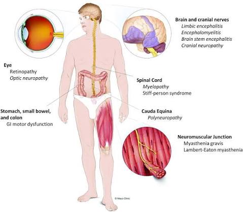
A neurological syndrome may be the first sign of cancer
Patients can develop autonomic dysfunction before, during or after the diagnosis of malignancy. The clinical course is usually subacute and progressive, evolving over weeks or months.
Orthostatic hypotension is often the most striking manifestation. In severe cases, the fall in blood pressure can be so profound that the patient becomes essentially bedridden despite aggressive attempts to maintain blood pressure.
But autonomic failure rarely limits itself to blood pressure.
A careful history may reveal:
- Constipation, nausea or severe gastrointestinal dysmotility
- Intestinal pseudo-obstruction
- Urinary retention or incontinence
- Erectile dysfunction
- Dry mouth and dry eyes
- Abnormal or absent sweating
- Pupillary abnormalities
- Cardiac tachyarrhythmias, bradycardia or even asystole
- Impaired heart-rate variability
- Exercise or heat intolerance
Peripheral and central neurological involvement may coexist. Sensory neuronopathy can produce painful, asymmetric or patchy sensory loss, while involvement of the central nervous system may lead to encephalitis, seizures, cognitive impairment, ataxia or altered consciousness.
In other words, the autonomic nervous system may be only one part of a much larger neurological syndrome.
Not every autonomic neuropathy is paraneoplastic
Autonomic failure has a broad differential diagnosis. Diabetes, amyloidosis, autoimmune diseases, neurodegenerative disorders, metabolic disorders, toxic neuropathies and certain infections can all produce similar manifestations.
The suspicion of a paraneoplastic syndrome increases when the dysfunction:
- develops rapidly or subacutely;
- is severe or multifocal;
- is accompanied by sensory neuronopathy or central neurological manifestations;
- occurs in association with a known cancer;
- or appears without an obvious explanation, particularly in a patient with risk factors for malignancy.
The challenge is that the tumor may still be invisible when the neurological disease begins.
Small-cell lung cancer is the classic association, but it is far from the only one. Thymoma, lymphomas, breast and ovarian cancers, and a variety of gastrointestinal, genitourinary and other malignancies have all been associated with paraneoplastic neurological syndromes.
What is happening to the nervous system?
The underlying mechanism is an immune response directed against onconeural antigens.
Some tumors express proteins that resemble, or are identical to, proteins normally found in neurons. The immune system recognizes these tumor-associated antigens and mounts an immune response. Unfortunately, that response can extend to healthy neural tissue.
This is why the neurological syndrome is not caused by direct tumor invasion.
Classical paraneoplastic antibodies are often directed against intracellular neuronal proteins. In these cases, the antibodies themselves may be more useful as markers of an underlying cytotoxic T-cell-mediated immune response than as the direct cause of neuronal injury.
Other antibodies target proteins exposed on the neuronal cell membrane and appear to be more directly pathogenic. These include antibodies against the ganglionic acetylcholine receptor and voltage-gated calcium channels.
Histologically, the process may produce neuronal loss accompanied by inflammatory infiltrates and perivascular lymphocytic inflammation.
Paraneoplastic autonomic neuropathy associated with anti-Hu antibodies. Histological sections demonstrate inflammatory mononuclear infiltrates and neuronal degeneration involving the central nervous system in a patient with autonomic failure and small-cell lung carcinoma.
Image: H&E staining. The original description demonstrates inflammatory infiltrates involving the inferior olive, hippocampus, midbrain and thoracic spinal cord.
The antibodies: useful clues, but not all the story
Several neuronal antibodies have been associated with paraneoplastic autonomic dysfunction.
Anti-ganglionic acetylcholine receptor antibodies
Antibodies against the α3 subunit of the ganglionic nicotinic acetylcholine receptor (gAChR) are strongly associated with autoimmune autonomic ganglionopathy.
They interfere with transmission in sympathetic and parasympathetic ganglia and can produce widespread autonomic failure.
Only a minority of autoimmune autonomic ganglionopathy cases are paraneoplastic. Reported associated malignancies include small-cell lung cancer, thymoma, bladder cancer and rectal carcinoma.
Importantly, these antibodies target ganglionic acetylcholine receptors and should not be confused with antibodies against the muscle acetylcholine receptor seen in myasthenia gravis.
Anti-Hu (ANNA-1)
Anti-Hu is one of the most important antibodies in paraneoplastic neurological disease and has a particularly strong association with small-cell lung cancer.
Its neurological manifestations are diverse because Hu antigens are expressed throughout the nervous system. Sensory neuronopathy is characteristic, but autonomic dysfunction is common enough to be an important clinical clue.
Gastrointestinal dysmotility is particularly frequent.
Anti-Hu positivity should therefore prompt a search for an underlying malignancy even when the initial presentation appears predominantly neurological or autonomic.
Anti-NMDA receptor antibodies
Anti-NMDA receptor encephalitis is better known for its psychiatric and neurological manifestations—psychosis, seizures, memory disturbance and altered consciousness—but autonomic instability can be prominent.
The association with ovarian teratoma is particularly important, although other tumors have occasionally been implicated.
Severe autonomic instability may include cardiac rhythm disturbances and, rarely, cardiac arrest.
Anti-VGCC antibodies and Lambert-Eaton syndrome
In Lambert-Eaton myasthenic syndrome (LEMS), antibodies against P/Q-type voltage-gated calcium channels impair neurotransmitter release at the neuromuscular junction.
The result is predominantly proximal muscle weakness, classically accompanied by reduced reflexes and autonomic symptoms.
Because these channels are also involved in autonomic neurotransmission, patients may experience dry mouth, constipation, erectile dysfunction and other manifestations of autonomic dysfunction.
Approximately half or more of LEMS cases are associated with malignancy, most commonly small-cell lung cancer.
Unlike many other paraneoplastic neurological syndromes, the autonomic symptoms of LEMS are often relatively mild and may improve substantially when the underlying cancer is treated.
Other antibodies
The list continues to grow.
CRMP5/CV2, PCA-2, CASPR2, VGKC-complex antibodies, amphiphysin, GAD65 and several other antibodies have been associated with neurological syndromes that may include autonomic dysfunction.
The important clinical point is that antibody testing is not simply a search for a single “paraneoplastic antibody.”
Different antibodies may produce overlapping syndromes, and more than one antibody can occasionally be detected in the same patient.
Conversely, a patient can have a convincing paraneoplastic neurological syndrome with no currently identifiable antibody.
A negative antibody panel therefore does not automatically exclude the diagnosis.
How do we diagnose it?
The diagnosis requires the integration of three elements:
clinical phenotype + antibody profile + evidence of cancer.
The diagnostic framework for paraneoplastic neurological syndromes was substantially revised in 2021 by an international panel of experts.
Rather than simply dividing syndromes into “classical” and “non-classical,” the updated criteria classify neurological phenotypes according to their risk of an underlying cancer, while antibodies are categorized according to their association with malignancy.
The system also introduced the PNS-Care Score, which incorporates:
- the neurological phenotype;
- the antibody findings;
- the presence of cancer.
Scores range from 0 to 10:
- 8–10: definite PNS
- 6–7: probable PNS
- 4–5: possible PNS
- 0–3: non-PNS
This approach improves diagnostic consistency and reduces the risk of overdiagnosing paraneoplastic neurological disease based solely on antibody positivity.
Looking for the hidden tumor
When no malignancy is known, the investigation should not stop after a negative initial scan.
Because small-cell lung cancer is such an important association, chest imaging is an essential first step, particularly in patients with relevant risk factors.
If conventional imaging fails to identify a tumor but clinical suspicion remains high, FDG-PET/CT can increase the likelihood of detecting an occult malignancy.
Depending on the clinical context, investigations may extend to the abdomen and pelvis, brain imaging, mammography or gynecological assessment, and testicular ultrasound.
And surveillance matters.
If the initial cancer search is negative, repeated screening may be necessary because the neurological syndrome can precede the radiological detection of the tumor.
The neurological work-up
Serum testing should generally include an appropriate paraneoplastic antibody panel, rather than testing for a single antibody in isolation.
Cerebrospinal fluid can provide additional evidence of an inflammatory process. Findings may include:
- lymphocytic pleocytosis;
- elevated protein;
- oligoclonal bands;
- and, in some cases, paraneoplastic antibodies.
CSF cytology is particularly relevant when alternative diagnoses such as leptomeningeal carcinomatosis need to be excluded.
Electromyography and nerve-conduction studies can demonstrate peripheral neuropathy or disorders of neuromuscular transmission such as LEMS.
Formal autonomic testing—including heart-rate variability, autonomic reflex testing and sudomotor studies—can document autonomic dysfunction, particularly when the clinical manifestations are subtle.
Treatment: treat the cancer, protect the nervous system
There are two therapeutic targets:
the malignancy and the autoimmune process.
The most important intervention is treatment of the underlying cancer.
If the tumor can be removed or effectively controlled, progression of the neurological syndrome may stop and, in some patients, neurological function may improve.
Unfortunately, established neuronal destruction is often irreversible.
Immunotherapy is considerably less predictable. Corticosteroids, intravenous immunoglobulin, plasma exchange and other immunomodulatory strategies have been used, but their effectiveness varies greatly between syndromes.
This difference is important.
Antibodies directed against intracellular antigens such as Hu are often markers of a predominantly T-cell-mediated neuronal injury. Once neurons have been destroyed, eliminating the circulating antibody may not restore neurological function.
By contrast, disorders caused by antibodies directed against cell-surface proteins—such as the ganglionic acetylcholine receptor or P/Q-type calcium channels—may respond more readily to immunotherapy.
Specific syndromes therefore require specific therapeutic strategies.
Managing autonomic failure
While the underlying cancer and immune process are being treated, supportive management can be crucial.
For orthostatic hypotension:
- review and discontinue blood-pressure-lowering drugs when appropriate;
- increase fluid and salt intake when not contraindicated;
- use compression garments or abdominal binders;
- elevate the head of the bed;
- avoid prolonged recumbency;
- and change position slowly.
Patients with impaired sweating require particular attention to heat exposure and dehydration, as thermoregulation may be severely compromised.
Gastrointestinal dysmotility may require dietary modification, treatment of constipation and, in severe cases, management of intestinal pseudo-obstruction.
Urinary retention may require intermittent or indwelling catheterization.
And cardiac autonomic dysfunction deserves particular attention because severe dysautonomia can occasionally result in life-threatening bradyarrhythmias or asystole.
A rare syndrome with an important message
Paraneoplastic autonomic neuropathy is rare, but its clinical implications are significant.
The key message is not simply that cancer can cause autonomic dysfunction.
It is that an unexplained, rapidly progressive autonomic syndrome may be the neurological presentation of an occult malignancy.
The combination of severe autonomic failure, sensory neuronopathy, gastrointestinal dysmotility or other neurological abnormalities should therefore prompt clinicians to think beyond the autonomic nervous system itself.
Sometimes, the nervous system is telling us that the tumor is there—before we can see it.
Key points for the Emergency Physician
Paraneoplastic autonomic neuropathy is uncommon, but it can present to the Emergency Department before the underlying malignancy has been diagnosed. Recognizing the pattern may therefore be the first step toward identifying an occult cancer.

Think beyond dehydration
Severe orthostatic hypotension without an obvious explanation should not automatically be attributed to dehydration, medication or simple volume depletion.
Ask whether there are other signs of autonomic dysfunction: impaired sweating, dry mouth, pupillary abnormalities, urinary retention, constipation, erectile dysfunction or abnormal heart-rate responses.

Look for the neurological combination
The combination of autonomic failure + sensory neuronopathy + gastrointestinal dysmotility should raise particular suspicion for a paraneoplastic process.
The presence of encephalopathy, seizures, ataxia, brainstem signs or rapidly progressive neurological deficits should further increase the level of concern.

Small-cell lung cancer is the classic association
When paraneoplastic autonomic dysfunction is suspected, actively consider small-cell lung cancer, particularly in patients with relevant smoking history or other risk factors.
But do not stop there: thymoma, lymphoma, breast and ovarian cancers, and several gastrointestinal and genitourinary malignancies have also been associated with paraneoplastic neurological syndromes.

A negative antibody panel does not exclude PNS
Paraneoplastic antibodies are extremely useful diagnostic clues, but they are not a perfect diagnostic test.
Some patients with a convincing paraneoplastic neurological phenotype have no currently identifiable antibody. Conversely, antibody positivity must always be interpreted in the context of the clinical syndrome and cancer risk.

Dysautonomia can be life-threatening
Severe autonomic dysfunction may produce profound hypotension, bradycardia, tachyarrhythmias or, rarely, asystole.
Continuous cardiovascular monitoring may therefore be appropriate in patients with severe dysautonomia, particularly when there is unexplained haemodynamic instability.

Don’t forget thermoregulation
Anhidrosis can be clinically important. Patients may be unable to dissipate heat appropriately and are at risk of hyperthermia, particularly during physical exertion or exposure to high temperatures.

The cancer may not be visible yet
A normal initial CT does not necessarily end the investigation.
If the neurological phenotype and antibody profile strongly suggest a paraneoplastic syndrome, further oncological investigation—including FDG-PET/CT when appropriate—and repeated surveillance may be required.

Treat the cause, but don’t neglect the syndrome
The cornerstone of management is identification and treatment of the underlying malignancy. Neurological damage may stop progressing when the tumor is controlled, although established neuronal loss can be irreversible.
At the same time, aggressive management of autonomic complications is essential: haemodynamic support, fluid and electrolyte management, temperature control, gastrointestinal and bladder care, and appropriate cardiac monitoring.

The emergency department may be where the diagnosis begins
Perhaps the most important lesson is this:
«When autonomic failure is severe, unexplained and rapidly progressive, think about cancer—even when there is no known cancer.»
The autonomic nervous system may be the first system to reveal a malignancy that has not yet declared itself.
